
Mouse Secondary Antibodies

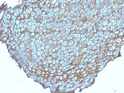
Biotium Desmoglein-3 (Squamous Cell Marker)(DSG3/2838), CF568 conjugate, 0.1mg/mL
Recognizes a protein of 130 kDa, identified as Desmoglein-3 (DSG3). This MAb is highly specific to Desmoglein-3 and does not cross-react with other members of the Desmoglein-family. DSG3 is a calcium-binding transmembrane glycoprotein component of desmosomes in vertebrate epithelial cells. Research has shown that DSG3 has a very high sensitivity (80%) and specificity (100%) in recognizing squamous cell carcinoma (SqCC). Therefore, DSG3 is considered a very important marker for lung SqCC and can be a useful ancillary marker to separate SqCC from other subtypes of lung cancer. Moreover, studies have shown that DSG3 expression in lung SqCC may indicate a poor prognosis. Primary antibodies are available purified, or with a selection of fluorescent CF Dyes and other labels. CF Dyes offer exceptional brightness and photostability. Note: Conjugates of blue fluorescent dyes like CF405S and CF405M are not recommended for detecting low abundance targets, because blue dyes have low

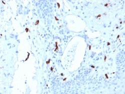
Biotium Tryptase (Mast Cell Marker) (TPSAB1/1961), CF568 conjugate, 0.1mg/mL
Tryptases comprise a family of trypsin-like serine proteases (peptidase family S1). Tryptases are stored in mast cell secretory granules and basophils. Mast cells are connective tissue cells derived from blood-forming tissues that line arterial walls and secrete substances, which mediate inflammatory and immune responses. Tryptases are released into the extracellular environment and are resistant to all known endogenous proteinase inhibitors. This antibody reacts with mast cells distributed in skin, synovium, lung, and heart. This antibody does not bind with any other cell type. Human mast cell tryptase is considered to be an important marker of mast cell activation and is an important mediator of inflammation. Mastocytosis is a term collectively used for a group of disorders in which there is abnormal accumulation of mast cells in one or multiple organs. Anti-tryptase, combined with anti-CD2, anti-CD25, and anti-CD117, can be useful in identifying reactive mast cell hyperpla
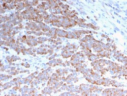
Biotium Cytokeratin 18 (KRT18)(rKRT18/1190), Biotin conjugate, 0.1mg/mL
This MAb reacts with a wide variety of simple epithelia. It does not react with stratified squamous epithelia. It reacts with epithelial tumors of the gastrointestinal tract, lung, breast, pancreas, ovary, and thyroid. Cytokeratin 18, which belongs to the type A (acidic) subfamily of low molecular weight keratins, exists in combination with cytokeratin 8. It was reported that tissues from gastrointestinal tract are positive for both cytokeratin 8 and 18 but do not contain cytokeratin 14. Tissues from gastrointestinal tract, respiratory tract and urogenital tract, as well as endocrine and exocrine tissues and mesothelial cells are positive for cytokeratin 18. Primary antibodies are available purified, or with a selection of fluorescent CF Dyes and other labels. CF Dyes offer exceptional brightness and photostability. Note: Conjugates of blue fluorescent dyes like CF405S and CF405M are not recommended for detecting low abundance targets, because blue dyes have lower fluore
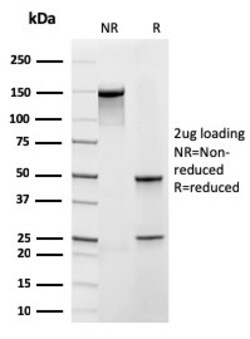
Biotium Frataxin (Mitochondrial Marker)(rFXN/2124), 0.2mg/mL
Frataxin is a nuclear-encoded mitochondrial protein implicated in Friedreich's ataxia (FRDA), a human autosomal recessive neurodegenerative disease. Lack of Frataxin causes iron to accumulate in the mitochondrial matrix suggesting that Frataxin is involved in mitochondrial iron homeostasis and possibly in iron transport. Frataxin has an alpha-beta fold consisting of two helices flanking an antiparallel beta sheet. Primary antibodies are available purified, or with a selection of fluorescent CF Dyes and other labels. CF Dyes offer exceptional brightness and photostability. Note: Conjugates of blue fluorescent dyes like CF405S and CF405M are not recommended for detecting low abundance targets, because blue dyes have lower fluorescence and can give higher non-specific background than other dye colors.
Biotium CD29 (Stem Cell Marker)(12G10), CF740 conjugate, 0.1mg/mL
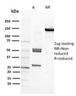
Recognizes CD29. Binds to the cell membrane of T- and B- lymphocytes, monocytes, platelets, fibroblasts, endothelial cells, mast cells and smooth muscle cells. CD29 is an adhesion receptor and co-stimulatory molecule. This antigen was initially characterized as GPIIa on platelets and as the common b1 subunit of the very late antigen (VLA) protein family. CD29 forms a non-covalent heterodimeric complex with integrin alpha subunits. It also mediates adhesion to invasion and thrombospondin. Primary antibodies are available purified, or with a selection of fluorescent CF Dyes and other labels. CF Dyes offer exceptional brightness and photostability. Note: Conjugates of blue fluorescent dyes like CF405S and CF405M are not recommended for detecting low abundance targets, because blue dyes have lower fluorescence and can give higher non-specific background than other dye colors.
Biotium C1QB / Complement C1q B-Chain(C1QB/2966), 0.2mg/mL
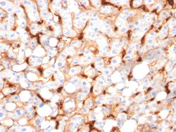
Complement C1q B Chain (C1qb) is a subunit of C1q. C1q is composed of 6 polypeptide chains each of C1qa, C1qb, and C1qc. C1q is a component of the classical complement pathway, and thereby plays an important role in the immune response. Each C1q subunit contains an N-terminal collagen-like region and a C-terminal C1q globular domain. The presence of receptors for C1q on effector cells modulates its activity, which may be antibody-dependent or independent. Macrophages are the primary source of C1q, while anti-inflammatory drugs as well as cytokines differentially regulate expression of the mRNA as well as the protein. C1q deficiency is associated with lupus erythematosus and glomerulonephritis. Primary antibodies are available purified, or with a selection of fluorescent CF Dyes and other labels. CF Dyes offer exceptional brightness and photostability. Note: Conjugates of blue fluorescent dyes like CF405S and CF405M are not recommended for detecting low abundance targets,

Biotium Protocadherin FAT2 (FAT Atypical Cadherin 2) (8C5), CF568 conjugate, 0.1mg/mL
This antibody recognizes a protein of 480 kDa, which is identified as FAT2. The cadherins represent a family of Ca2 -dependent adhesion molecules that function to mediate cell-to-cell binding that is critical for the maintenance of structure and morphogenesis. Cadherins each contain a large extracellular domain at the N-terminus, which is characterized by a series of five homologous repeats, the most distal of which is thought to be responsible for binding specificity. The relatively short C-terminal intracellular domain interacts with a variety of cytoplasmic proteins, including beta-catenin, to regulate cadherin function. The cadherin superfamily includes cadherins, protocadherins, desmogleins and desmocollins. FAT2 (FAT tumor suppressor homolog 2) is a single-pass type I membrane protein that belongs to the protocadherin subfamily of cadherins. FAT2 contains one Laminin G-like domain, two EGF-like domains and 32 cadherin domains and is believed to function as a cell adhesi
Biotium Crystallin Alpha B(CPTC-CRYAB-1), CF568 conjugate, 0.1mg/mL
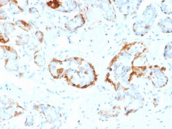
Crystallins are the major proteins of the vertebrate eye lens, where they maintain the transparency and refractive index of the lens. Crystallins are divided into B-crystallin is associated with many neurological diseases, and a missense mutation in this gene has co-segregated in a family with a Desmin-related myopathy. Primary antibodies are available purified, or with a selection of fluorescent CF Dyes and other labels. CF Dyes offer exceptional brightness and photostability. Note: Conjugates of blue fluorescent dyes like CF405S and CF405M are not recommended for detecting low abundance targets, because blue dyes have lower fluorescence and can give higher non-specific background than other dye colors.
Biotium DOG-1 / TMEM16A (Gastrointestinal Stromal Tumor Marker)(DG1/2831R), CF488A conjugate, 0.1mg/mL
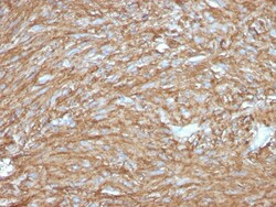
Expression of DOG-1 protein is elevated in the gastrointestinal stromal tumors (GIST's), c-kit signaling-driven mesenchymal tumors of the GI tract. DOG-1 is rarely expressed in other soft tissue tumors, which, due to appearance, may be difficult to diagnose. Immunoreactivity for DOG-1 has been reported in 97. 8 percent of scorable GIST's, including all c-kit negative GIST's. Overexpression of DOG-1 has been suggested to aid in the identification of GISTs, including Platelet-Derived Growth Factor Receptor Alpha mutants that fail to express c-kit antigen. The overall sensitivity of DOG1 and c-kit in GIST's is nearly identical: 94. 4% vs. 94. 7%. Primary antibodies are available purified, or with a selection of fluorescent CF Dyes and other labels. CF Dyes offer exceptional brightness and photostability. Note: Conjugates of blue fluorescent dyes like CF405S and CF405M are not recommended for detecting low abundance targets, because blue dyes have lower fluorescence and can
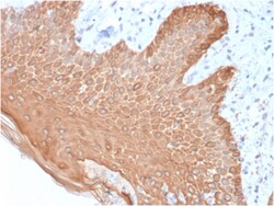
Biotium Cytokeratin, Multi (Epithelial Marker)(rKRT/457), Biotin conjugate, 0.1mg/mL
Twenty human keratins are resolved with two-dimensional gel electrophoresis into acidic (pI 6. 0) subfamilies. This antibody recognizes acidic (Type I or LMW) and basic (Type II or HMW) cytokeratins, including 59 kDa (CK4); 58 kDa (CK5); 56 kDa (CK6); 52 kDa (CK8); 56. 5 kDa (CK10); 53 kDa (CK13) and 45 kDa (CK18). This is a broad-spectrum antibody, which has been reported to differentiate epithelial tumors from non-epithelial tumors. Many studies have shown the usefulness of keratins as markers in cancer research and tumor diagnosis. Primary antibodies are available purified, or with a selection of fluorescent CF Dyes and other labels. CF Dyes offer exceptional brightness and photostability. Note: Conjugates of blue fluorescent dyes like CF405S and CF405M are not recommended for detecting low abundance targets, because blue dyes have lower fluorescence and can give higher non-specific background than other dye colors.
Biotium Cytokeratin 6 (KRT6) (Hyperproliferation-Related Keratin) (KRT6/1702), CF488A conjugate, 0.1mg/mL
This MAb recognizes a protein of 56 kDa, identified as cytokeratin 6 (CK6). In humans, multiple isoforms of Cytokeratin 6 (6A-6F), encoded by several highly homologous genes, have distinct tissue expression patterns, and Cytokeratin 6A is the dominant form in epithelial tissue. The gene encoding human Cytokeratin 6A maps to chromosome 12q13, and mutations in this gene are linked to several inheritable hair and skin pathologies. Keratins 6 and 16 are expressed in keratinocytes, which are undergoing rapid turnover in the suprabasal region (also known as hyper-proliferation-related keratins). Keratin 6 is found in hair follicles, suprabasal cells of a variety of internal stratified epithelia, in epidermis, in both normal and hyper-proliferative situations. Epidermal injury results in activation of keratinocytes, which express CK6 and CK16. CK6 is strongly expressed in about 75% of head and neck squamous cell carcinomas. Expression of CK6 is particularly associated with different
Biotium Cytokeratin 8 (KRT8)(B22.1), 1mg/mL
Cytokeratin 8 (CK8) belongs to the type II (or B or basic) subfamily of high molecular weight cytokeratins and exists in combination with cytokeratin 18 (CK18). CK8 is primarily found in the non-squamous epithelia and is present in majority of adenocarcinomas and ductal carcinomas. It is absent in squamous cell carcinomas. Hepatocellular carcinomas are defined by the use of antibodies that recognize only cytokeratin 8 and 18. CK8 exists on several types of normal and neoplastic epithelia, including many ductal and glandular epithelia such as colon, stomach, small intestine, trachea, and esophagus as well as in transitional epithelium. Anti-CK8 does not react with skeletal muscle or nerve cells. Epithelioid sarcoma, chordoma, and adamantinoma show strong positivity corresponding to that of simple epithelia (with antibodies against CK8, CK18 and CK19). Reportedly, anti-CK8 is useful for the differentiation of lobular ('ring-like, perinuclear') from ductal ('peripheral-predomina

Biotium Villin (GI-Mucosal & Urogenital Brush Border Marker) (VIL1/1314), CF488A conjugate, 0.1mg/mL
This antibody recognizes a protein of 95 kDa, which is identified as villin. It is a major constituent in the microvilli, which compose the brush border of epithelial cells forming absorptive surfaces of the intestinal and renal proximal tubular epithelia. Anti-Villin labels the brush border area in the gastrointestinal mucosal epithelium and urogenital tract. Among neoplasms, villin is predominantly expressed in tumors of colorectal origin. Antibody to villin is useful in identifying malignant cells from primary and metastatic colorectal carcinomas. This antibody also labels Merkel cells of the skin.Primary antibodies are available purified, or with a selection of fluorescent CF Dyes and other labels. CF Dyes offer exceptional brightness and photostability. Note: Conjugates of blue fluorescent dyes like CF405S and CF405M are not recommended for detecting low abundance targets, because blue dyes have lower fluorescence and can give higher non-specific background than oth
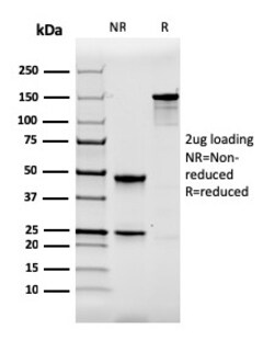
Biotium Calbindin 1 (CALB1)(CALB1/2364), 1mg/mL
Calbindin 1, also known as calbindin D28K, is a member of a family of calcium binding proteins. This protein family also includes calmodulin, calbindin 2, S-100, and troponin C. Calbindin 1 is a 261-amino acid protein with 6 EF-hand domains, 4 of which are active calcium-binding domains. Expressed in brain, ovary, uterus, testis, pancreas, liver, kidney and intestine, calbindin 1 acts as a calcium-buffering agent and alters the activity of the plasma membrane ATPase. In neuronal cells, calbindin 1 modulates calcium channel activity, calcium transients and intrinsic neuronal firing activity. Also, calbindin 1 has been implicated to play a role in apoptosis and microtubule function. Primary antibodies are available purified, or with a selection of fluorescent CF Dyes and other labels. CF Dyes offer exceptional brightness and photostability. Note: Conjugates of blue fluorescent dyes like CF405S and CF405M are not recommended for detecting low abundance targets, because blue dyes
Biotium Cytokeratin 7(K72.7), CF740 conjugate, 0.1mg/mL
This antibody recognizes an intermediate filament protein (IFP) of 55 kDa, which is identified as cytokeratin 7. This MAb is highly specific to cytokeratin 7 and shows no cross-reaction with other IFPs. Cytokeratin 7 is a basic cytokeratin, which is found in most glandular and transitional epithelia but not in the stratified squamous epithelia. Keratin 7 is expressed in the epithelial cells of ovary, lung, and breast but not of colon, prostate, or gastrointestinal tract. This MAb is highly useful in distinguishing ovarian carcinomas (keratin 7 ) from colon carcinomas (keratin 7-).Primary antibodies are available purified, or with a selection of fluorescent CF Dyes and other labels. CF Dyes offer exceptional brightness and photostability. Note: Conjugates of blue fluorescent dyes like CF405S and CF405M are not recommended for detecting low abundance targets, because blue dyes have lower fluorescence and can give higher non-specific background than other dye colors.